Mole mapping
Each nevus (commonly referred to as a mole), indicates a benign tumor of the epidermis that appears as a flat (macula) or a slightly elevated (papula) elementary skin lesion.
The term nevus is erroneously used to also indicate skin neoformations, including angiomas, fibroids and various other lesions. This is why, in the scientific terminology, the word “nevus” is followed by an adjective that better defines it.
If the nevus originates from the melanocyte (skin cells responsible for the production of melanin) then we refer to a melanocytic nevus; if instead it originates from keratinocytes, then it’s an epidermal nevus. Again, if it originates from cells constituting the skin appendages (in general, cells that derive from the ectoderm), then we are referring to dermal nevi, sebaceous nevi, verrucous nevi, follicular nevi and so on.
Congenital nevi (i.e. present at birth) are considered hamartomas; acquired nevi are instead considered benign neoplasms.
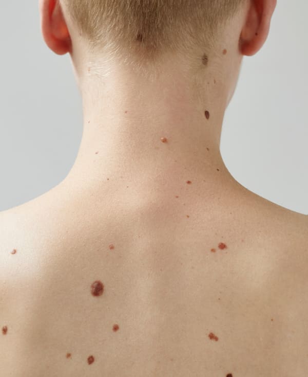
Regardless of the type, however, moles represent a skin abnormality that can be found anywhere on the body, even on the head, on the soles of the feet or in the eyes. However, they are completely physiological: most people have between 10 and 50 of them. As mentioned, some of these nevi are present at birth, others appear over time.
Benign moles are usually small – no more than half a centimeter wide – and have a rounded shape, with defined contours. Their color ranges from pink to dark brown. Most benign moles on the skin are completely harmless. A very small part of them, however, can degenerate and give rise to a specific aggressive skin tumor: melanoma. This transformation can occur spontaneously, even if it is proven that external stresses such as exposure to UV rays tend to favor it.
Suspicious moles, such as those that create an aesthetic problem for their shape and dimensions, must be removed. It is essential to refer to a reconstructive surgeon with oncological experience (also when removing benign moles). At Image Regenerative Clinic, Professor Carlo Tremolada places his renowned ability in this regard at the patient’s disposal, because moles often represent a cause for apprehension, or embarrassment – or both.
For this precise reason, when a nevus arouses suspicion, Prof. Tremolada performs an excision followed by a histological examination, in order to give the patient a safe evaluation.
It is always advisable to periodically undergo a mapping of the moles to verify their size and any increase in that, the type and – after some time – the onset of new moles.
There are several techniques for removal: the surgical one remains the most appropriate for extirpating the root and preventing the mole from reforming. The round block suture technique, invented by Professor Tremolada himself, makes it possible to minimize scarring and have the possibility of intervening again with confidence in the event of any surprises revealed by the histological examination.
«Today the only valid reason not to remove a mole is that the mole itself is neither suspicious nor active, and it is also an appreciable distinctive personal mark the patient particularly likes. Another reason not to intervene, if the mole is not dangerous, is that the future scar may be unsightlier than the mole itself.»
Prof. Carlo Tremolada
© Istituto Image 2024